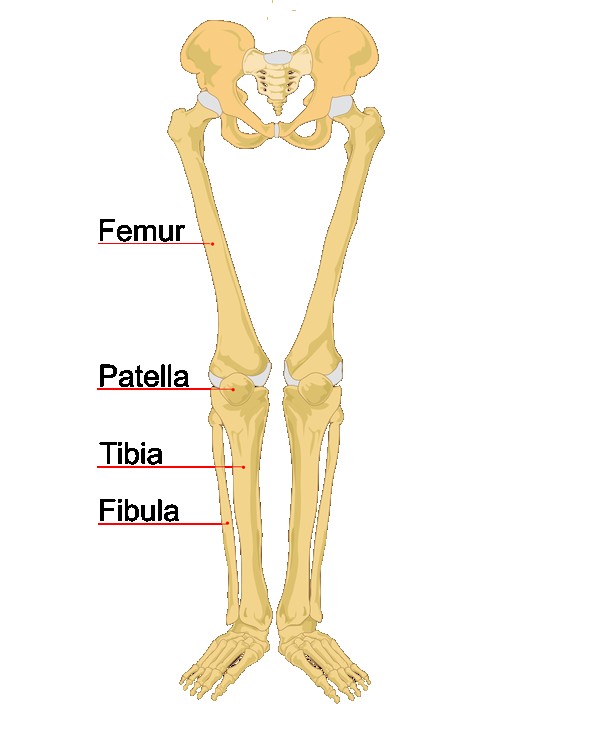
Leg Bone Diagram / Comparison of bones of forearm and lower leg - anterior ... / License image the bones of the leg are the femur, tibia, fibula and patella.
Leg Bone Diagram / Comparison of bones of forearm and lower leg - anterior ... / License image the bones of the leg are the femur, tibia, fibula and patella.. Femur bone diagram get rid of wiring diagram problem. Joints of hand anterior view, lateral view, right hand. The humerus and the femur are corresponding bones of the arms and legs, respectively. Learn vocabulary, terms and more with flashcards, games and other study tools. Bone tissue, also called osseous tissue, is classified as either compact bone.
Master leg and knee anatomy using our topic page. The human leg, in the general word sense, is the entire lower limb of the human body, including the foot, thigh and even the hip or gluteal region. Click now to learn more about the bones, muscles, and soft tissues tibia: When you stand or walk, all the weight of your upper body rests on them. The axial skeleton and the appendicular formed by the left and right hip bones, the pelvic girdle connects the lower limb (leg) bones to the axial.
A basic human skeleton is studied in schools with a simple ... from i.pinimg.com These bones are arranged into two major divisions: Time to jump right into the biggest and strongest bones in the human body. Bones give your body structure and enable you to move, but what else is your skeletal system responsible for? Click now to learn more about the bones, muscles, and soft tissues tibia: Leg bone anatomy diagram diagram of human leg human anatomy. Human skeleton long bones of arms and legs britannica. Spongy bone is composed of trabeculae that contain the bones of the pelvis, skull, spine, and legs are the most commonly affected. The foot bones shown in this diagram are the talus, navicular, cuneiform, cuboid, metatarsals and calcaneus.
Human skeleton long bones of arms and legs britannica.
Pngtree offers bone diagram png and vector images, as well as transparant background bone diagram clipart images and psd files. Femur bone diagram get rid of wiring diagram problem. Spongy bone is composed of trabeculae that contain the bones of the pelvis, skull, spine, and legs are the most commonly affected. The foot bones shown in this diagram are the talus, navicular, cuneiform, cuboid, metatarsals and calcaneus. The humerus and the femur are corresponding bones of the arms and legs, respectively. The foot bones shown in this diagram are the talus, navicular, cuneiform, cuboid, metatarsals. Cheek bone (zygoma) upper jaw (maxilla). Joints of hand anterior view, lateral view, right hand. Master leg and knee anatomy using our topic page. Explore the fascination world of human bones. Human skeleton long bones of arms and legs britannica. Click now to learn more about the bones, muscles, and soft tissues tibia: Learn how to draw the femur, patella, tibia, and fibula in this lesson!
12 photos of the diagram of leg bones. Your legs are two of your most important body parts. The largest and most medial leg bone, forming both the knee and ankle joints. The red bone marrow inside of bones produces most of the blood cells, including erythrocytes (red blood cells), leukocytes (white blood cells), and thrombocytes (platelets). The foot bones shown in this diagram are the talus, navicular, cuneiform, cuboid, metatarsals.
Leg & Foot Archives - Medical Art Library from medicalartlibrary.com In the leg, the interosseous membrane extends between the tibia and the fibula, running along the crests of the bones. Your leg bones are the longest and strongest bones in your body. Each leg is made up of four bones. Explore the fascination world of human bones. Lower jaw (mandible) collar bone. Download the free graphic resources in the form of png, eps. Cheek bone (zygoma) upper jaw (maxilla). The axial skeleton and the appendicular formed by the left and right hip bones, the pelvic girdle connects the lower limb (leg) bones to the axial.
Human skeleton long bones of arms and legs britannica.
Explore the fascination world of human bones. Master leg and knee anatomy using our topic page. The red bone marrow inside of bones produces most of the blood cells, including erythrocytes (red blood cells), leukocytes (white blood cells), and thrombocytes (platelets). Download 2,751 bone diagram stock illustrations, vectors & clipart for free or amazingly low rates! These bones are arranged into two major divisions: The bones of the leg are the femur, tibia, fibula and patella. They allow you to move and provide support for your upper body. 12 photos of the diagram of leg bones. The human leg, in the general word sense, is the entire lower limb of the human body, including the foot, thigh and even the hip or gluteal region. The humerus and the femur are corresponding bones of the arms and legs, respectively. Your legs are two of your most important body parts. While their parts are similar in general, their structure has been adapted to differing functions. Bones of the leg and foot, lower leg bone anatomy, leg bones anatomy, leg muscles, leg bones diagram, leg bone structure, leg anatomy muscles, parts of the lower leg.
Download 2,751 bone diagram stock illustrations, vectors & clipart for free or amazingly low rates! Visit kenhub for more skeletal system quizzes. These bones are arranged into two major divisions: Pngtree offers bone diagram png and vector images, as well as transparant background bone diagram clipart images and psd files. The largest and most medial leg bone, forming both the knee and ankle joints.
human-leg-bones-labeled_l | Biolulia European Sections from bioluliaes.files.wordpress.com They allow you to move and provide support for your upper body. Cheek bone (zygoma) upper jaw (maxilla). This bright worksheet helps your child bring these technical terms down to size. Click now to learn more about the bones, muscles, and soft tissues tibia: Leg bone anatomy diagram diagram of human leg human anatomy. However, the definition in human anatomy refers only to the section of the lower limb extending from the knee to. The largest and most medial leg bone, forming both the knee and ankle joints. Your leg bones are the longest and strongest bones in your body.
Disposition of rotator cuff muscles diagram.
Cheek bone (zygoma) upper jaw (maxilla). Master leg and knee anatomy using our topic page. You'll learn about the muscles, bones, and other structures of each area of the leg. Your leg bones are the longest and strongest bones in your body. New users enjoy 60% off. In the leg, the interosseous membrane extends between the tibia and the fibula, running along the crests of the bones. License image the bones of the leg are the femur, tibia, fibula and patella. The largest and most medial leg bone, forming both the knee and ankle joints. While their parts are similar in general, their structure has been adapted to differing functions. Download 2,751 bone diagram stock illustrations, vectors & clipart for free or amazingly low rates! Time to jump right into the biggest and strongest bones in the human body. Pngtree offers bone diagram png and vector images, as well as transparant background bone diagram clipart images and psd files. Disposition of rotator cuff muscles diagram.